Dermacademy Challenge – Clinical Case #3 – Pr. Zaslavsky
- 30min

- 1/2
- 2/2
Clinical case shared by Pr. Zaslavsky
Clinical Presentation
Patient K., 5 years old, accompanied by her mother N., 36 years old, was referred for consultation. According to the patient's mother at admission, the patient complained of skin eruptions on the body, upper and lower limbs, neck, and scalp.The child has had this condition for about a year when an inflammation site first appeared on the back of the neck. The patient's mother does not associate the disease onset with anything.
Family History
The patient's mother has had cutaneous tuberculosis since early childhood. Negative allergic anamnesis. No bad habits. Past diseases: acute respiratory disease. Denies sexually transmitted diseases, hepatitis, HIV. No history of hemotransfusions or surgeries. Mother's pregnancy and childbirth without abnormalities.
Physical Examination
A coarse keloid scar formed on the surgery site, and a spot, growing in size, appeared on the head. Later, the skin process began to spread: skin eruptions appeared on the body and limbs, as well as on the scalp. The mother began self-administration of rifampicin lotion on the head.
Satisfactory general condition. Regular build. Clear regular heart sounds. Vesicular breath sounds heard throughout all lung fields, without rales. Abdomen is soft and nontender, nondistended, non-asymmetric. The liver edge does not protrude below the costal margin; dense, elastic consistency; nontender, even. The spleen is not palpable. The kidneys are nontender on palpation. Murphy's punch sign is negative on both sides. Lymph nodes are not enlarged. Stool and urination without abnormalities.
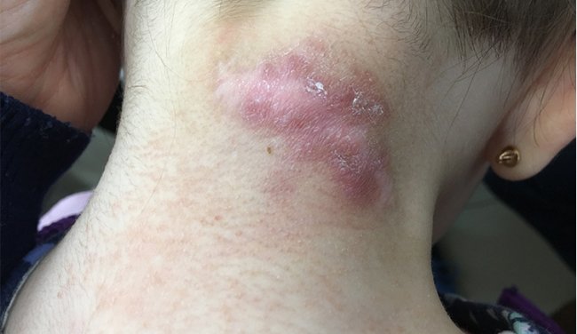
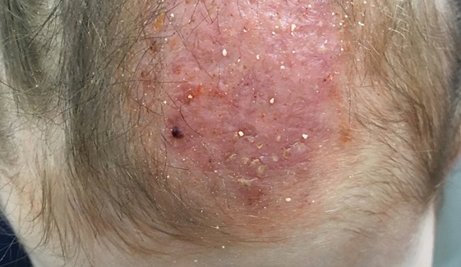
Multiple eruptions, predominantly located on the scalp, back of the neck, side surfaces of the body, upper and lower limbs. An inflammatory lesion on the scalp, 6x4 cm, with signs of atrophy, pustular rash elements, round-shaped, of scarlet-brown color (Figure 1). Multiple dense conical-shaped tubercles of golden-brown color covered with easily removable crusts (Figure 2) are noted on the skin of upper and lower limbs. Single, slightly tender dense mulberry nodules, of 1.5–2 cm, surgical scars (Figure 3) are noted on the back surface of the neck. The nodules are slightly tender on palpation; diascopy reveals a strong positive ""apple-jelly"" sign. Peripheral lymph node groups are not enlarged, not adherent to the skin, movable, nontender. Nail plates on fingers and toes are unchanged. No subjective perception.
Laboratory Result
In recent years, Mantoux tuberculin skin tests (2TU) have shown a normergic reaction with augmentation (р = 15 mm). Inactivity of TB infection, absence of clinical and radiological evidence suggestive of TB, negative reactions to the test with Diaskintest and ELISPOT required consultation by an oncologist who sampled material for histological verification of diagnosis. Pathologists suggested granulomatous inflammation.
Biopsy Result
According to the description of the morphological material, there are tissue fragments covered with non-keratinized stratified squamous epithelium with diffuse round-cell infiltrate in the subepithelial stroma. Pronounced caseous necrosis with epithelioid cell granulomas and single Langhans multinucleated giant cells, mainly without a clear demarcation area (Figure 4), is identified in the infiltrate. Single granulomas are surrounded by a thin rim of collagen fibers. Non-keratinized stratified squamous epithelium with diffuse infiltration by segmented neutrophils and mononuclear leukocytes. The molecular genetic test of the biopsy material has isolated the DNA of the M.tuberculosis complex which allows for verification of cutaneous tuberculosis diagnosis.
Diagnosis
Based on the complaints, medical history data, results of ELISPOT, positive PCR-based diagnostics, and the morphological pattern, the diagnosis of cutaneous tuberculosis (scrofuloderma, lichenoid tuberculosis, lupus vulgaris) has been verified.
Management and outcome
Patient was sent to phthisiatrician (tuberculotherapist).
Teaching points
Cutaneous tuberculosis is a clinically and morphologically heterogeneous group of skin diseases directly or indirectly caused by mycobacteria of the tuberculosis complex.Cutaneous tuberculos is occupying the 5th place among all localizations of extra-pulmonary tuberculosis.
Manifestations of cutaneous tuberculosis are extremely diverse and depend on the immune status and ways of penetration of mycobacteria into the skin. Skin involvement can occur as a result of exogenous inoculation, contiguous spread from an adjacent focus, or hematogenous spread from other foci. Family cases of disseminated cutaneous tuberculosis are described.
Bibliography
- Van Vooren JP, Schepers K: screening for tuberculosis. Rev Med Brux 2013;34:301-305.
- World health oganisation (WHO) Global tuberculosis control: WHO report 2013; Genva: WHO 2013.
- O.B.Boudghene StambouliA.Dib Lachachi La tuberculose cutanée : toujours un problème d’actualité et de santé publique en Algérie : à propos de 7 observations récentes Ann Dermatol Vénéréol 2018;145 :A32.
- Diagnostic Standards and Classification of Tuberculosis in Adults and Children. This official statement of the American Thoracic Society and the Centers for Disease Control and Prevention was adopted by the ATS Board of Directors, July 1999. This statement was endorsed by the Council of the Infectious Disease Society of America, September 1999.Am J Respir Crit Care Med. 2000 Apr;161(4 Pt 1):1376-95.