Rebeca, 41 years old: Polymorphous light eruption.
 Oct. 2021
Clinical case shared by Pr. Zaslavsky
Clinical Presentation
A female patient, 16 years old, with complaints of skin eruptions on the body and limbs was admitted to the Department of Dermatovenerology, St. Petersburg State Pediatric Medical University. The patient has had this condition since the age of 5, when, according to her parents, the spots first appeared on lower limbs. When referring to the local dermatologist in Syktyvkar, the patient was diagnosed with various disorders: Devergie's disease, atopic dermatitis, allergic dermatitis, diffuse eczema, toxidermia. The patient was treated with topical steroids, nonspecific desensitizing, detoxification; however, there was no significant improvement. At a later stage, the course of skin lesions became torpid: very slowly, spots resolved spontaneously after several years, but then they appeared again either at the sites of pre-existing eruptions or on visually healthy skin. In the following years, the disease progressed: spots similar to previously existing ones started appearing on the body, upper and lower limbs, and their number increased significantly.
Physical Examination
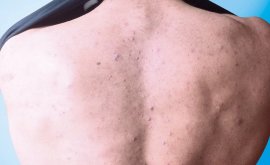
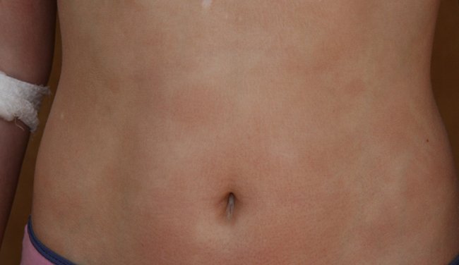
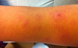
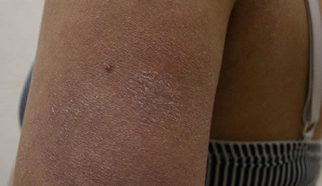
The skin lesion process is generalized. Unaffected skin areas are preserved only on the face, palms, and soles. Spots of irregular shape and of various sizes, 5 to 20 cm in area, are noted over the entire skin cover. The spots merge with each other forming large foci covering whole anatomical regions. Most spots are pink, although they vary in color in different areas: from light-brown to dark red-brown with a bluish tint. The spots on the inner surface of the hip are more intense and infiltrated. Skin phototype II. Lymph nodes are not enlarged. The patient's condition is satisfactory.
Laboratory Result
The results of complete blood count and biochemical blood test are within normal.
Biopsy Result
Histological and immunohistochemical findings Micro: a skin area with orthokeratosis, parakeratosis, irregular acanthosis in the epidermis. Single pathologic mitoses and the accumulation of atypical lymphocytes (Pautrier's microabscess) are occasionally found in the spinous layer. Intraepithelial lymphocytes are also found. Prominent stripe-like infiltrate consisting mainly of atypical lymphocytes of triangular shape and cerebriform nuclei is revealed in the derma. This infiltrate is separated from the epidermis with collagen fibers. Atypical cells tend to penetrate the epidermis. Atypical lymphocytes in the epidermis are of larger sizes than in the derma (Figure 3, 4). The immunohistochemical assay reveals a pronounced expression of T-lymphocytes CD2+, CD3+, СD4+, CD5+ marker both in the epidermis and the derma, while no expression of cytotoxic T-lymphocyte CD8 marker is revealed.
Diagnosis
Taking into account immunohistochemical findings, the described histological change may be observed in mycosis fungoides. Mycosis fungoides is classified Т2b, N0, M0, B0.
Management and outcome
Based on recommendations for treatments of primary skin lymphomas, PUVA therapy was recommended to patient. However, taking into the account age of patient, small amount of infiltrated long persistent spots and absence of photosensitizer, recommended for classical methods of phototherapy (psoralen), it was indicated:
After 30 phototherapy procedures, clinical response of disease significantly decreased. During morphological examination only signs of autopathic inflammation appeared were found.
Teaching points
Clinical and morphological heterogeneity of malignant lymphoproliferative lesions of the skin is explained by the peculiarities of their pathogenesis and organization of the lymphoid tissue in the skin. These features determine the authenticity of therapeutic approaches to patients with this pathology and exclude the use of standard regimens of polychemotherapy used in oncology due to their low efficacy.
Mycosis fungoides is the most common form of cutaneous T-cell lymphoma. Its annual incidence is between 1/350,000 and 1/110,000. The male to female ratio is 2:1. Mycosis fungoides affects more adults and elderly people. The onset of the condition manifests through spots and/or patches on the skin, usually asymmetrical and localised on the buttocks and areas protected from sun exposure (lower torso, thighs, and in women, breasts). If the scalp is affected, alopecia may develop.
Rare cases of paediatric MF have been reported: The median age upon diagnosis was 11 years old.The condition is generally indolent despite persisting into adulthood in a large proportion of these paediatric cases.
Bibliography