Cutaneous small vessel vasculitis
Cutaneous vasculitis with erythema multiforme-like target appearance following trifluridine/tipiracil treatment for colon adenocarcinoma
Symptoms/Signs
Physical examination revealed a rash of circular erythematous lesions with clear margins bilaterally on the lower limbs, some confluent on the pretibial surfaces, with
a central vesicobullous formation. Some lesions had a target-like appearance.
Patient photographs
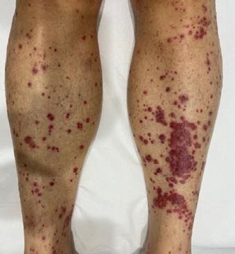
Clinical presentation
Physical examination revealed a rash of circular erythematous lesions with clear margins bilaterally on the lower limbs, some confluent on the pretibial surfaces, with a central vesicobullous formation. Some lesions had a target-like appearance.
Medical history
In September 2020, he was diagnosed with grade 3 adenocarcinoma of the ascending colon with metastases in the lymph nodes, liver and lungs.
Previous lines of treatment: capecitabine;
FOLFOX combination (FOLinic acid, Fluorouracil, OXaliplatin) + bevacizumab;
FOLFIRI combination (FOLinic acid, Fluorouracil, IRInotecan) + bevacizumab.
In the month prior to the visit, he underwent a course of antineoplastic treatment with a combination of trifluridine and tipiracil hydrochloride (a nucleoside analogue of thymidine and a thymidine phosphorylase inhibitor, respectively).
Differential diagnosis
Drug-induced cutaneous vasculitis of the small vessels
Urticaria
Drug-induced rash
Erythema multiforme
Stevens Johnson syndrome
Diagnostic tests
Clinical examination of the entire skin area helps identify the differential diagnoses. Erythema multiforme is usually triggered by a viral infection (HSV), is rarely drug-induced, and predominantly affects the upper limbs and face. In urticaria, pomphoid lesions show no signs of epidermal damage at their centre and are typically temporary. Drug-induced rash has fewer elements on the skin. Stevens Johnson syndrome is a rare life-threatening cutaneous adverse drug reaction characterised by skin and mucosal epidermal detachment and systemic involvement.
The general diagnostic framework for skin vasculitis includes laboratory testing of ESR, complement, and autoantibodies, as well as diagnostic skin biopsy to confirm the presence of leukocytoclastic vasculitis.
Description of the disease
Drug-induced cutaneous small vessel vasculitis (CSVV) are predominant in the extremities, such as the lower limbs, and occurs with eruptive lesions 1 to 3 weeks after taking the implicated drug. The pathogenic mechanism is mediated by type II (cytotoxicity) or type III (immune complex) immune reactions.
Lesions often appear as purpuric macules or erythematous or urticarial papules; target lesions are sometimes observed.
Injuries may be asymptomatic or associated with itching and burning. Internal organ involvement, which is usually rare, should be ruled out; discontinuation of the responsible drug usually leads to resolution. Post-inflammatory hyperpigmentation is a normal finding after healing.
Join our community to read the full article
L'Oréal Dermatological Beauty Pro is a digital community empowering healthcare professionals to improve their daily practice of dermatology through cutting edge research, science and education on skin and hair care.
Sign Up For Free TodayAlready registered? Log in